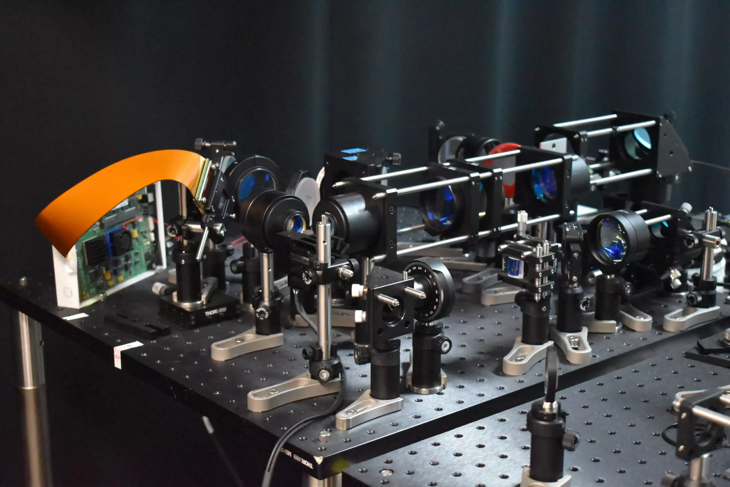
The field of neuroscience has seen a groundbreaking development with the creation of a new two-photon fluorescence microscope that is capable of capturing high-speed images of neural activity at cellular resolution. This innovative approach presents a significant improvement over traditional two-photon microscopy methods, offering researchers a clearer view of how neurons communicate in real-time. The implications of this advancement are vast, potentially leading to new insights into brain function and neurological diseases.
The key feature of this new microscope is the incorporation of a novel adaptive sampling scheme that differs from the traditional point illumination used in conventional two-photon microscopy. By utilizing line illumination, researchers are able to image neuronal activity in a mouse cortex at speeds ten times faster than before, while also reducing the amount of laser power on the brain tissue by more than tenfold. This allows for a more efficient and less damaging imaging process, making it ideal for studying the dynamics of neural networks in real-time.
One of the most promising aspects of this new technology is its potential to aid in the study of neurological diseases at their earliest stages. By observing neural activity in real-time, researchers can gain valuable insights into the pathology of conditions such as Alzheimer’s, Parkinson’s, and epilepsy. This deeper understanding could pave the way for more effective treatments and interventions in the future.
Traditional two-photon microscopy, while capable of providing detailed images, has been hindered by its slow imaging speed and potential for harm to brain tissue. The new sampling strategy introduced by the researchers addresses these limitations by using a line of light to illuminate specific areas of the brain where neurons are active. This adaptive sampling technique allows for a larger area to be imaged at once, speeding up the process while minimizing the risk of damage to the tissue.
The researchers achieved this breakthrough by employing a digital micromirror device (DMD) to dynamically shape and steer the light beam, ensuring precise targeting of active neurons. This innovative approach not only enhances the speed and accuracy of imaging but also reduces the total light energy deposited into the brain tissue. Additionally, the researchers developed a technique to mimic high-resolution point scanning, enabling the reconstruction of high-quality images from fast scans.
Moving forward, the researchers plan to further enhance the capabilities of the microscope by integrating voltage imaging for a rapid readout of neural activity. This expanded functionality will allow for a more comprehensive understanding of neural interactions and the functional architecture of the brain. Real neuroscience applications, such as observing neural activity during learning and studying brain activity in disease states, are also on the horizon. Moreover, efforts to improve the user-friendliness and portability of the microscope aim to make it more accessible for research in the field of neuroscience.
The development of this new two-photon fluorescence microscope represents a significant milestone in the field of neural imaging. By enabling high-speed imaging of neuronal activity with reduced risk to living tissue, this technology has the potential to revolutionize our understanding of brain function and neurological diseases. Through continued innovation and application in real-world research settings, this advancement promises to open up new opportunities for studying dynamic neural processes in unprecedented detail.


Leave a Reply